A Photographic Atlas for the Anatomy and Physiology Laboratory 7th Edition PDF Download
Book Details
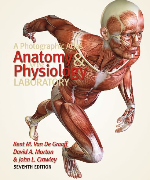
Human anatomy is the scientific discipline that investigates the structure of the body and human physiology is the scientific discipline that investigates how body structures function. These subjects may be taught independent of each other in separate courses, or they may be taught together in integrated anatomy and physiology courses. Regardless of whether or not anatomy is taught independently from physiology or if the two disciplines are integrated as a single course, it is necessary for a student to have a conceptualized visualization of body structure and a knowledge of its basic descriptive anatomical terminology in order to understand how the body functions. A Photographic Atlas for the Anatomy and Physiology Laboratory is designed for all students taking separate or integrated courses in human anatomy and physiology. This atlas can accompany and will augment any human anatomy, human physiology, or combined human anatomy and physiology textbook. It is designed to be of particular value to students in a laboratory situation and could either accompany a laboratory manual or in certain courses, serve as the laboratory manual. Anatomy and physiology are visually oriented sciences. Great care has gone into the preparation of this photographic atlas to provide students with a complete set of photographs for each of the human body systems. Human cadavers have been carefully dissected and photographs taken that clearly depict each of the principal organs from each of the body systems. Cat dissection, fetal pig dissection, and rat dissection are also included for those students who have the opportunity to do similar dissection as part of their laboratory requirement. In addition, photographs of a sheep heart dissection are also included. A visual balance is achieved in this atlas between the various levels available to observe the structure of the body. Microscopic anatomy is presented by photomicrographs at the light microscope level and electron micrography from scanning and transmission electron microscopy. Carefully selected photographs are used throughout the atlas to provide a balanced perspective of the gross anatomy.
More Info
Year:2011
Edition:7
Publisher:Morton Publishing Company
Language:english
Pages:224 / 226
ISBN 10:0895828758
ISBN 13:9780895828750
File:PDF, 19.37 MB
Download
Socia Drive
