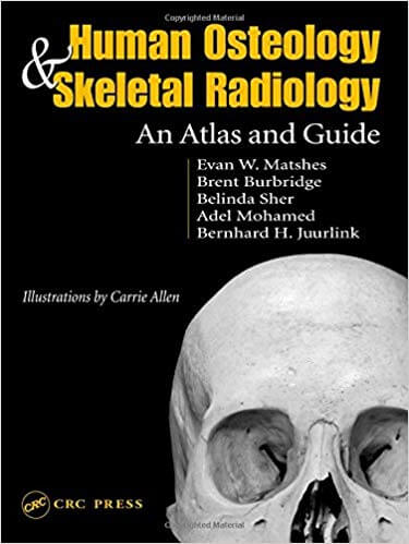
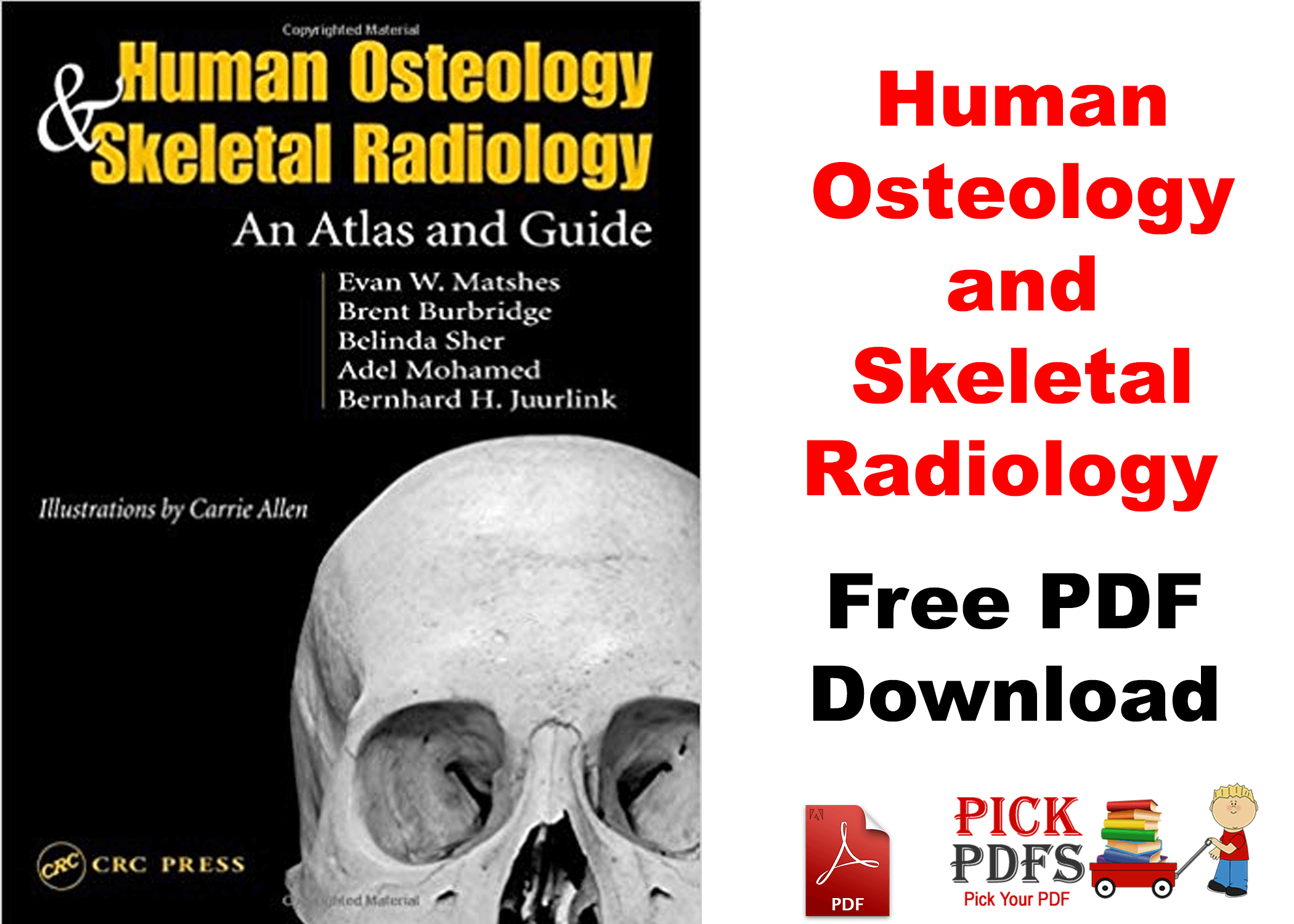
Human Osteology and Skeletal Radiology An Atlas and Guide PDF download
Features
Human Osteology and Skeletal Radiology PDF
An Atlas and Guide features nearly 700 photographs, line drawings, and radiographs demonstrating individual bones, or collections of bones, from both a distant perspective and more detailed angles. This atlas of skeletal anatomy covers general and specific anatomic terms, includes comparative images of bones in photographic and radiographic form to aid in recognition, and notes important comparisons among adult, juvenile, and fetal bones. It discusses each bone on an individual basis and describes how to “side” bones and identify fragments.
Intended as a field guide for investigations and a lab guide in gross anatomy and skeletal specimen studies, this atlas provides easy and rapid identification of bone material. It takes you far beyond the bare bones of anatomy to aid in skeletal recognition in any situation.
Description
The incorporation of a radiological aspect with the traditional photographic and graphic approaches is long overdue and makes this a valuable addition to the reference library of any skeletal or forensic anthropologist.” Thomas D. Holland, Ph.D., Scientific Director, Joint POW/MIA Accounting Command Central Identification Laboratory, Hickam Air Force Base, Hawai’i, USA “provides readers with a user-friendly and hands-on approach to identifying and siding all elements of the human skeleton. One advantage of this text over others is its clarity of skeletal landmarks as depicted in large photographs. Another advantage is the layout of the chapters in a commonsense way that minimizes the ‘seek and find’ approach common to many texts. I will certainly add this to my personal library and use it as a required text in my forensic anthropology class. combines all of the essential elements in one text.
User Reviews for Human Osteology and Skeletal Radiology An Atlas and Guide
A classic in its field, Human Osteology has been used by students and professionals for nearly two decades. Now revised and updated for a third edition, the book continues to build on its foundation of detailed photographs and practical real-world application of science. New information, expanded coverage of existing chapters, and additional supportive photographs keep this book current and valuable for both classroom and field work.
Osteologists, archaeologists, anatomists, forensic scientists, and paleontologists will all find practical information on accurately identifying, recovering, and analysing and reporting on human skeletal remains and on making correct deductions from those remains.
About the Author
Bernhard H. Juurlink is Professor and Head, Department of Anatomy and Cell Biology, College of Medicine, University of Saskatchewan.
Table of Contents
Human Osteology and Skeletal Radiology An Atlas and Guide includes the following units, sections, and chapters:
UNIT 1: THE AXIAL SKELETON
- Anatomical Terms of Direction and Osteologic Terminology
- The Skull
- The Hyoid and Spine
- The Sternum and Ribs
UNIT 2: THE APPENDICULAR SKELETON
- The Shoulder and Upper Limbs
- The Pelvis
- The Upper Extremities
UNIT 3: WRAPPING IT UP
- References and Index
- File Size: 16 MBs
- 448 Pages
- English Language
- 3-Star Rating
Below is a Download Button for Human Osteology and Skeletal Radiology An Atlas and Guide PDF Download

Disclaimer:
This site complies with DMCA Digital Copyright Laws. Please bear in mind that we do not own copyrights to this book/software. We’re sharing this with our audience ONLY for educational purposes and we highly encourage our visitors to purchase the original licensed software/Books. If someone with copyrights wants us to remove this software/Book, please contact us. immediately.
You may send an email to taimourdev@gmail.com for all DMCA / Removal Requests
![Pastest Essential Revision Notes For MRCP PDF 4th Edition [Direct Links]](https://pickpdfs.com/wp-content/uploads/2019/07/MRCP2-768x549.png)
