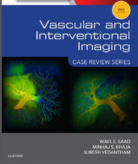
Vascular and Interventional Imaging Case Review Series 3rd Edition PDF »

Book Description:

Increase your knowledge and improve your image interpretation skills using the proven and popular Case Review approach! Drs. Wael E. Saad, Minhaj S. Khaja, and Suresh Vedantham have updated this new edition to include nearly 200 clinically relevant cases with associated images, multiple-choice questions, answers, and rationales – all organized by level of difficulty and designed to reinforce your understanding of the essential principles needed to interpret a wide range of vascular and interventional images.

Key Features

  • Study effectively with content that addresses contemporary medical practice emphasizing physics, clinical evaluation, and treatment following The Joint Commission and the Accreditation Council for Graduate Medical Education.
  • View cases that reflect contemporary practice, thanks to new, multi-modality imaging, including color images and 3D reconstructions.
  • Stay up to date with recent innovations in vascular and interventional radiology with a more diverse case selection, updated procedural techniques and inventory, and new and updated references from reputable sources.
  • Get fresh perspectives from nearly 200 updated or new cases reflecting the most recent changes in vascular and interventional radiology, including portal hypertension, aortic disease, interventional oncology, peripheral arterial disease, and complex venous disease.

Table of Contents:

Opening Round
Fair Game
Challenge
Supplemental Figures
Index of Cases
Index of Terms

Vascular and Interventional Imaging Case Review Series 3rd Edition PDF Free Download

About the Authors/Authors Notes :

Wael Saad Author
Affiliations and Expertise
Professor of Radiology, Director, Vascular & Interventional Radiology, Director, Neurointerventional Radiology, Department of Radiology, University of Michigan Medical Center, Ann Arbor, Michigan

Minhaj Khaja Author
Affiliations and Expertise
Assistant Professor of Radiology, Vascular & Interventional Radiology, Department of Radiology, University of Michigan Medical Center, Ann Arbor, Michigan

Suresh Vedantham Author
Affiliations and Expertise
Professor of Radiology & Surgery, Mallinckrodt Institute of Radiology, Washington University School of Medicine, St Louis, Missouri

More Info and Download Links:

No. of pages: 649
Language: English
File Size : 210 MB
File Format : PDF

Download Links For the Book : Vascular and Interventional Imaging Case Review Series 3rd Edition PDF

Taybi and Lachman’s Radiology of Syndromes Metabolic Disorders and Skeletal Dysplasias 5th Edition PDF »

Preface:

It has been over 30 years since the publication of the first edition of this book in 1975. Over the years there has been amazing progress in the fields of medical imaging, syndromology, metabolic disorders, and skeletal dysplasias. Even more recently the explosion in the field of molecular biology and gene identification has made the subjects covered in this edition even more exciting. I have tried to incorporate as much of this into the text as possible. The first edition of the book covered about 540 entities. In the second edition, about 700 subjects were discussed. By the third edition nearly 1000 syndromes, metabolic disorders, and skeletal dysplasias were included. The fourth edition contained over 1000 fully described entities as well as 1250 entries in abbreviated form included in a gamuts section. The gamuts references numbered about 1800. The third and fourth editions were divided into three separate chapters (Syndromes, Metabolic Disorders, and Skeletal Dysplasias). For the reader’s convenience, I have chosen to abridge this edition into two main sections with both syndromes and metabolic disorders alphabetically in one section and the skeletal dysplasias in a separate section.
I hope this will make usage of the text easier. This fifth edition contains 120 new fully described entries, including both entities and tables. There are about 90 new syndromes and metabolic disorders and 35 new skeletal dysplasias. To make the text more user-friendly for the geneticist as well as the radiologist and clinician, I have included the OMIM number for appropriate entries. The gamuts section has also been significantly improved and expanded with the addition of a searching CD included with this edition to allow searches using several symptoms or signs to narrow the search to a smaller number of entities, whether they are common, uncommon, or rare. The gamuts section has been increased so that there are over 22,500 entries and over 4500 gamuts-related references. The fifth edition also contains some new tables, including the overall radiographic expression of the entire group of mucopolysaccharidoses/mucolipidoses (dysostosis multiplex) and the general radiographic expression of chromosome abnormalities. A number of new tables regarding groups of important mutational disorders are also found. In this edition the articles are cited not only by the author’s name, but also the publication year, which will make it easier to find specific references. Appendix A contains the most recent Nosology and Classification of Genetic Disorders of Bone (Skeletal Dysplasias). In Appendix B, I have included a chapter on the radiographic approach to the skeletal dysplasias, which should be valuable to the novice as well as to those taking board examinations. I hope that these changes will make the text more userfriendly for all of us.

Taybi and Lachman’s Radiology of Syndromes Metabolic Disorders and Skeletal Dysplasias 5th Edition PDF Free Download, Taybi and Lachman’s Radiology of Syndromes Metabolic Disorders and Skeletal Dysplasias 5th Edition PDF Ebook Free

Review of Radiology 3rd Edition PDF download

Review of Radiology 3rd Edition PDF Free Download
Preface:

The IBQ section would definitely help the students in preparing for the different types of questions based in images expected to be asked in the examination. The online pool of questions is going to be a backbone for the preparation of IBQs in any examination not just for radiology but for other subjects too. Finally the Section-I) has a collection of important factual information of radiology including the investigations of choices, important systemic signs and important principles of radiology which might be helpful to solve questions from any clinical subject if understood rightly. How to Read this Book The best way to read this book is to read the theory given at the beginning of the chapter first and then solve the MCQs. The MCQs of a particular system are arranged randomly without mentioning the years. Absolute care has been taken to avoid repetition of the MCQs as lots of time is wasted in solving the same MCQ again and again. We want to stress on this fact that nowadays in the exams, same questions are not repeated but same topics are repeated and hence students are advised to focus more on the theoretical concepts rather than just MCQs. It is this reason that, deliberately years of the questions and superscript on important lines have not been put as we believe that each line written is a potential MCQ. We suggest that section A, C and 13 should be read together where section C should be read in associating with other clinical subjects like medicine, surgery, orthopedics, pediatrics, gynecology-obstetrics as they are interlinked. We wish that our students will go through this book thoroughly and will do excellent in their examinations. Best wishes for your postgraduate medical entrance examination. Your queries and feedback will always be welcomed. You are free to contact at email: reviewofradiologye gmail.com

More Info:

Keywords:
Review of Radiology 3rd Edition Free Download ,Review of Radiology 3rd Edition Ebook ,Review of Radiology 3rd Edition Free

Cancer Imaging Lung and Breast Carcinomas Volume 1 PDF download

Cancer Imaging Lung and Breast Carcinomas Volume 1 PDF Free Download
Preface:

The primary objective of this series, CANCER IMAGING, is to present the readers with the most up-to-date instrumentation, general applications, as well as specific applications of imaging technology to diagnose various cancers. The present work concentrates on the application of this technology to the diagnosis of lung and breast carcinomas, the two major worldwide cancers. In this work we discuss strategies for imaging cancer and clinical applications of this technology and explain the role of molecular imaging in early therapy monitoring. In addition, we cover the following topics: synthesis and use of different contrast agents, especially the most commonly used tracer, 18F-fluorodeoxyglucose (FDG), in conjunction with imaging modalities (e.g., magnetic resonance imaging [MRI]); the advantages and limitations of the tracers; and rational and protocol details of whole-body screening with computed tomography (CT), positron emission tomography (PET), and PET/CT. We also describe the role of imaging in drug development, gene therapy, and therapy with monoclonal antibodies; for example, we describe the advantages of preclinical immuno-PET. The advantages of hybrid modalities, such as PET/CT (e.g., in lung cancer), are also presented. The importance of the use of imaging for clinical diagnosis is presented in detail, and the relationship between radiation dose and image quality is discussed. In addition, we present detailed methods for absorbed X-ray dose measurement in mammography to avoid the risk of radiation-induced carcinogenesis. We also detail lung cancer screening, staging, and diagnosing, applying different imaging modalities and point out false-negative and false-positive images potentially encountered in various body parts when imaged. Imaging modalities, including high-resolution CT, thinsection CT, computer-aided diagnosis with CT, low-dose helical CT, and FDG-PET/CT, used for staging and diagnosing lung cancer (e.g., non-small cell lung carcinoma), are discussed. Details of a large number of imaging modalities, including optical mammography, digital mammography, contrast-enhanced MRI, SPECT, color-Doppler sonography, and sonography, used for diagnosing breast cancer are presented. Other topics include use of imaging for diagnosing invasive lobular carcinoma, density of breast carcinoma, axillary lymph node status in breast cancer, small-size primary breast cancer, and microcalcification in breast lesions.

More Info:

Print Length: 664 pages
1 edition (October 25, 2007)
Language: English

Keywords:
Cancer Imaging Lung and Breast Carcinomas Volume 1 Free Download ,Cancer Imaging Lung and Breast Carcinomas Volume 1 Ebook ,Cancer Imaging Lung and Breast Carcinomas Volume 1 Free

Physicians’ Cancer Chemotherapy Drug Manual 2019 PDF download

Physicians’ Cancer Chemotherapy Drug Manual 2019 PDF Free Download
Preface:

The development of effective drugs for the treatment of cancer represents a significant achievement beginning with the discovery of the antimetabolites and alkylating agents in the 1940s and 1950s. The success of that effort can be attributed in large measure to the close collaboration and interaction between basic scientists, synthetic organic chemists, pharmacologists, and clinicians. This tradition continues to flourish, especially as we now enter the world of pharmacogenomics, genomics, proteomics, and other omics science along with the rapid identification of new molecular targets for drug design and development. In this, our 19th edition, we have condensed and summarized a wealth of information on chemotherapeutic and biologic agents in current clinical practice into a reference guide that presents essential information in a practical and readable format. The primary indications, drug doses and schedules, toxicities, and special considerations for each agent have been expanded and revised to take into account new information that has been gathered over the past year. In this edition, we have included 13 new agents that have all been approved by the FDA within the past year and we have also updated new indications for previously approved agents. This drug manual is divided into five chapters. Chapter 1 gives a brief overview of the key principles of cancer chemotherapy and reviews the clinical settings where chemotherapy is used. Chapter 2 reviews individual chemotherapeutic and biologic agents that are in current clinical use; these agents are presented in alphabetical order according to their generic name. In this chapter, specific details are provided regarding drug classification and category, key mechanisms of action and resistance, critical aspects of clinical pharmacology and pharmacokinetics, clinical indications, special precautions and considerations, and toxicity

More Info:

Paperback: 694 pages
19 edition (December 21, 2018)
Language: English

Thyroid Ultrasonography and Fine Needle Aspiration Biopsy PDF download

Thyroid Ultrasonography and Fine Needle Aspiration Biopsy PDF Free Download
Preface:

Disorders of the thyroid and parathyroid glands are common and neck ultrasonography has stood the test of time and established itself as the primary imaging modality of not only the thyroid gland but also of the parathyroid glands and cervical lymph nodes. When clinically indicated, neck ultrasonography is invaluable and can be seen as an extension of the physical examination. It is performed in the office without any patient preparation and allows the physician to carry-out ultrasound-guided biopsies of anterior and lateral neck structures. Physicians who are not personally performing neck ultrasonography and ultrasound guidedprocedures still need to be quite familiar with the ultrasound features of the normal and abnormal thyroid gland, parathyroid glands and cervical lymph nodes. This is particularly important as there is a wide variation in the standards of neck ultrasonography reporting. The authors of this book are Endocrinologists with an interest in thyroid and parathyroid disorders, who have been performing neck ultrasonography and ultrasound-guided procedures of the neck for more than 15 years. When we set out to write this book, our aim was to provide a basic introduction to neck ultrasonography and to allow the reader to quickly search for relevant information about the ultrasound feature or condition in question. To aid with this task, we tried to include as many ultrasound images as possible and to limit the amount of text as we believe in the saying “a picture is worth a thousand words”. This book would be a useful tool for endocrinologists, endocrine surgeons, cytopathologists, ultrasonographers and other health care workers with an interest in thyroid and parathyroid disorders. We would like to thank our family and friends who supported us with the writing of this book and hope that you enjoy it!

More Info:

Print Length: 165 pages
Language: English

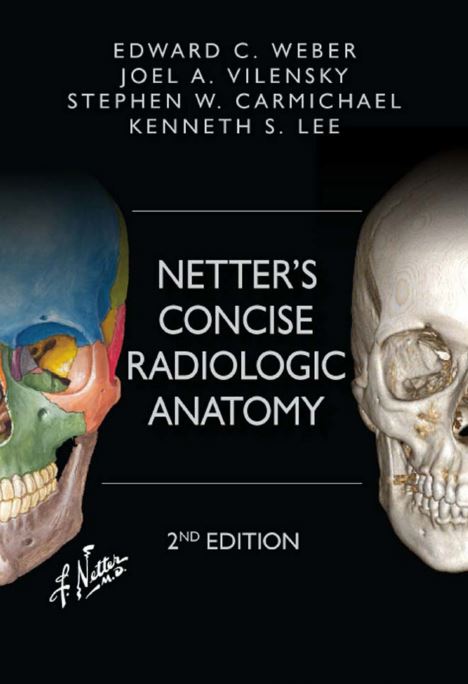
Netter’s Concise Radiologic Anatomy 2nd Edition PDF

Book Details

Netter’s Concise Radiologic Anatomy 2nd Edition PDF Free Download

Diagnostic medical images are now an integral component of contemporary courses in medical gross anatomy. This primarily reflects the steadily increasing teaching of clinical correlations within such courses. Accordingly, radiographic images are included in all gross anatomy atlases and textbooks. These images are typically plain radiographs, axial CT/MRI (computed tomography/magnetic resonance image) scans, and angiograms of various parts of the vascular system. Although such images reflect the capabilities of diagnostic imaging technology of perhaps 25 years ago, they do not reflect the full integration of computer graphics capabilities into radiology. This integration has resulted in a tremendous expansion in the ability of radiology to represent human anatomy. The active process of reformatting imaging data into optimal planes and types of image reconstruction that best illustrate anatomic/pathologic features is not limited to academic centers. To the contrary, the graphics workstation is now a commonly used tool in the practice of diagnostic radiology. Special views and image reconstructions are currently part of the diagnostic process and are usually made available to all those participating in patient care, along with an interpretation by the radiologist that describes the pathology and relevant anatomy. This situation led us to the realization that any student of anatomy would benefit from early exposure to the manner of appearance of key anatomic structures in diagnostic images, especially advanced CTs and MRIs. Thus, in 2007 we (a radiologist and two anatomists) chose to develop an atlas that illustrates how modern radiology portrays human anatomy. To accomplish this task, we decided to match modern diagnostic images with a subset of the anatomic drawings from the Atlas of Human Anatomy by Dr. Frank H. Netter. Netter’s atlas has become the gold standard of human anatomy atlases. Its images are quite familiar to the vast majority of students who complete a course in human gross anatomy.

More Info

Year:2014

Edition:2nd

Language:english

Pages:519 / 551

ISBN 13:978-1-4557-5323-9

File:PDF, 115.56 MB