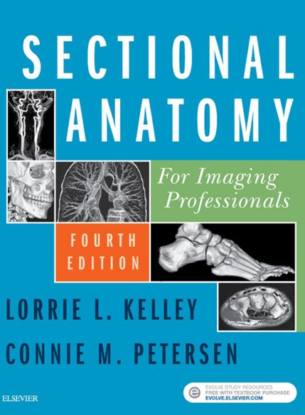
This text was written to address the needs of today’s practicing health professional. As technology in diagnostic imaging advances, so does the need to competently recognize and identify cross-sectional anatomy. Our goal was to create a clear, concise text that would demonstrate in an easy-to-use yet comprehensive format the anatomy the health professional is required to understand to optimize patient care. The text was purposely designed to be used both as a clinical reference manual and as an instructional text, either in a formal classroom environment or as a self-instructional volume. Included are close to 1000 high-quality MR and CT images for every feasible plane of anatomy most commonly imaged. An additional 350 anatomic maps and line drawings related to the MR and CT images add to the learner’s understanding of the anatomy being studied. In addition, pathology boxes describe common pathologies related to the anatomy presented, assisting the reader in making connections between the images in the text and common pathologies that will be encountered in clinical practice. Updated tables are used to summarize and organize key information in each chapter. For example, tables that summarize muscle group information include points of origin and insertion, as well as functions, for the muscle structures pertinent to the images the reader is studying. NEW TO THIS EDITION • Updated content to reflect the latest ARRT and ASRT curriculum guidelines • Expanded images in the lymphatic system • Second color added to the design to make difficult content easier to digest CONTENT AND ORGANIZATION The images include identification of vital anatomic structures to assist the health professional in locating and identifying the desired anatomy during actual clinical examinations. The narrative accompanying these images clearly and concisely describes the location and function of the anatomy in a format easily understood by health professionals. The text is divided into chapters by anatomic regions. Each chapter of the text contains an outline that provides an overview of the chapter’s contents, pathology boxes that briefly describe common pathologies related to the anatomy being presented, tables designed to organize and summarize the anatomy contained in the chapter, and reference illustrations that provide the correct orientation for ease of locating the anatomy of interest.

Sectional Anatomy for Imaging Professionals 4th Edition PDF
by Taimour
Taimour
Dr. Taimour is a dedicated medical professional and passionate advocate for international medical graduates seeking to pursue their dream of becoming a doctor abroad. With a wealth of experience and firsthand knowledge of the challenges and rewards of this journey, Dr. Harrison is committed to helping aspiring physicians navigate the complex world of medical licensure exams, such as the USMLE and PLAB, and find their path to success in foreign medical practice.